Traditional anatomy education has long been limited by practical constraints. The biggest challenge is the scarcity of donated bodies. Dissection is irreversible; once a mistake is made, it cannot be undone. Beginners often face psychological and ethical pressure when working with real cadavers.
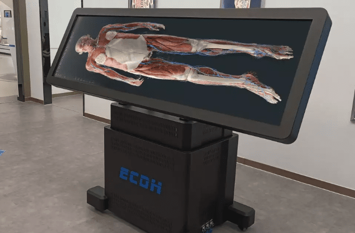
How can these challenges be addressed? The virtual dissection table offers an innovative solution.
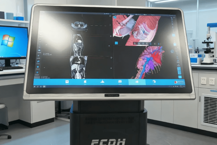
This is an integrated hardware and software platform that utilizes 3D technology to create interactive, high-resolution anatomical models. These 3D models enable users to explore human anatomy within a dynamic and immersive environment.
Challenge 1: Resource Scarcity and High Costs
One of the biggest obstacles in anatomy education is the limited availability of body donations. Medical schools often struggle to provide enough cadavers for all students. Even when available, cadavers involve high costs for transportation, preservation, and long-term maintenance.
The virtual dissection table addresses these issues effectively.
- Schools can invest in multiple devices instead of relying solely on donated bodies.
- Students can perform repeated dissections on realistic digital models, referred to as virtual cadavers, without worrying about damaging specimens. The models were derived from actual CT and MRI data.
- Virtual anatomy tables require almost no maintenance compared to traditional labs. The result is a cost-effective, sustainable, and scalable approach to anatomy education.
Challenge 2: Irreversible Operations and Risks
Dissecting a cadaver is an irreversible action. Students may hesitate to explore complex structures fully due to the fear of making mistakes. Beyond this, preservatives like formalin pose potential health risks, making repeated practice less safe.
By contrast, the virtual dissection table allows students to experiment freely.
- Features such as “undo” and “redo” functions let learners correct errors instantly.
- Multiple rehearsals are possible, enabling students to build confidence and competence.
- Touch control operation allows for cutting and observing structures at different levels of the human body from various directions and angles.
- No health hazards
Using a virtual anatomy table, learners can repeatedly explore intricate structures, improving their understanding of human anatomy safely and efficiently.
Challenge 3: Difficulty Understanding Complex Spatial Relationships
Understanding how organs, nerves, and vessels relate to each other in 3D space is a hard part of medicine. Traditional dissection is “superficial to deep.” Static 2D diagrams in textbooks also fail to capture the complexity of the human form.
Here, anatomy 3D technology shines.
- A virtual dissection table offers zoom, rotation, and layered dissection features, allowing students to examine every structure from multiple angles.
- Layer-by-layer exploration enhances spatial reasoning, helping learners understand how organs, muscles, and vessels relate in three-dimensional space.
Digital tools make complex structures more accessible. It’s valuable for students struggling with spatial visualization.
Challenge 4: Individual Learning Differences
Not all students progress at the same pace. Some may need extra time to grasp certain anatomical concepts, while others quickly advance through the curriculum. Once the lab session ends, access to the labs is usually restricted, leaving little room for independent review.
The virtual anatomy table supports self-paced learning.
- Students can repeat lessons, revisit challenging sections, and take quizzes at their own speed.
- Many systems, such as the Diguhuman virtual anatomy table, include a comprehensive test bank, helping reinforce knowledge and improve retention.
By personalizing the learning experience, these tables ensure that students achieve a comprehensive understanding of human anatomy, regardless of individual learning differences.
Digihuman’s Virtual Dissection Table
For institutions looking to modernize digital anatomy education, Digihuman virtual anatomy table is a standout solution.
It combines high-resolution anatomy 3D models, interactive tools, and a user-friendly interface suitable for beginners and advanced learners. The system functions as a true virtual cadaver, offering the benefits of traditional dissection without the limitations.
With Digihuman’s platform, students can explore every aspect of human anatomy, from bones and muscles to nerves and vessels. The virtual anatomy table supports repeated practice, enhances spatial understanding, and accommodates self-paced learning, all while reducing costs and eliminating health risks.
It’s an ideal tool for medical schools, clinical healthcare, or life science education.
Conclusion
Challenges in traditional anatomy education, such as scarce resources, irreversible operations, complex spatial relationships, and varying learning paces, can limit students’ learning outcomes.
Investing in a virtual dissection table is no longer just a supplement; it is a transformative step in modern medical education, bridging the gap between labs and the learning needs of today. If you have any needs, contact Digihuman now for more information!